Detaillierte Informationen: Fibrotische Erkrankungen

ImmuneNTech antifibrotische Therapien

Während viele chronische Krankheiten kausal behandelt werden können, z.B. durch die Eliminierung von Infektionen, immunsuppressive Therapien oder die Behandlung des metabolischen Syndroms, haben die Patienten bei der Erstdiagnose häufig bereits eine fortgeschrittene Fibrose, die als irreversibel gilt. Die meisten fibrotischen Erkrankungen können nicht wirksam durch eine kausale Therapie behandelt werden. Daher besteht ein dringender Bedarf an spezifischen Therapien zur Behandlung der Fibrose. Bislang ist jedoch noch kein wirksames antifibrotisches Medikament für den klinischen Einsatz zugelassen.

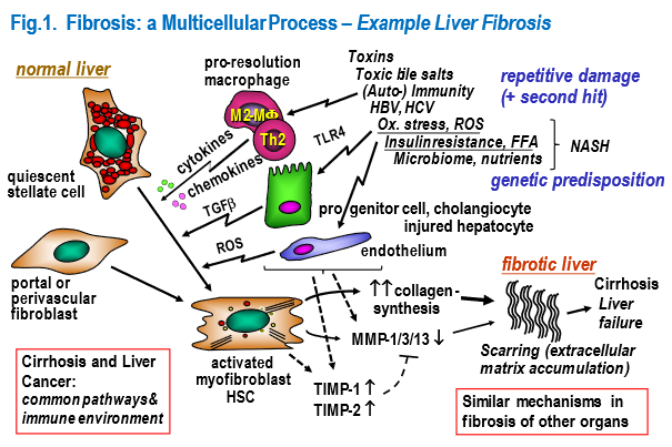
ImmuneNTech hat sich auf Schlüsselmechanismen und zelluläre Akteure konzentriert, die das Fortschreiten der Fibrose (Fibrogenese) oder auch die Umkehrung der Fibrose (Fibrolyse) vorantreiben, insbesondere in der Leber, aber auch in anderen Organen, in denen die Fibrose auf ähnliche Mechanismen und Zellen zurückzuführen ist. Zu den therapeutischen Wirkstoffen gehören kleine Wirkstoffmoleküle, interferierende RNA (siRNA) oder Peptide, welche spezifische Gene, Transkripte oder Proteine so modulieren, dass die Fibrogenese gehemmnt oder die Fibrolyse gefördert wird. Zu den Zielzellen gehören die ECM-produzierenden Effektorzellen (aktivierte (Myo)Fibroblasten), Myofibroblasten-aktivierende Zellen (epitheliale Vorläuferzellen) und Makrophagen, welche die Fibrogenese und Fibrolyse maßgeblich steuern (Abbildung 1).

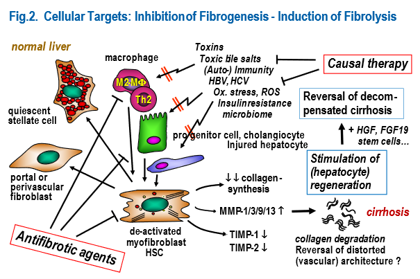
Unsere therapeutischen Ansätze basieren auf Molekülen, welche die involvierten Zellen im Sinne einer Hemmung der Fibroseprogression oder der Induktion einer Fibroseregression beeinflussen (Abbildung 2)

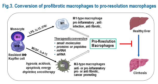
Die Wissenschaftler von ImmuneNTech haben mehrere Strategien entwickelt und validiert, die auf fibrogene Cholangiozyten, Myofibroblasten/Lebersteernzellen, Makrophagen und die ECM abzielen. Dabei wurden gut validierte Nagetiermodelle der Leberfibrose (parenchymatös, biliär und NASH) eingesetzt. Auf der Grundlage dieser Arbeiten sind einige der so getesteten Arzneimittel bereits in klinischen Studien der Phase 2 (siehe unten aufgeführte Schlüsselpublikationen). Ein Beispiel für unsere neuen Strategien zur Beeinflussung von Makrophagen bei Leberfibrose findet sich in Abbildung 3.

  1. Fraser D, Wang XY, Lund J, Nikolic N, Iruarrizaga-Lejarreta M, Skjaeret T, Alonso C, Kastelein JJP, Rustan C,  Kim YO, Schuppan D. A structurally engineered fatty acid, icosabutate, suppresses liver inflammation and fibrosis in NASH. J Hepatol. 2021; Dec 13; Epub ahead; doi: 10.1016/j.jhep.2021.11.015.
  2. Wagner WL, Föhst S, Hock J, Kim YO, Popov Y, Schuppan D, Schladitz K, Redenbach C, Ackermann M. 3D analysis of microvasculature in murine liver fibrosis models using synchrotron radiation-based microtomography. Angiogenesis. 2021;24:57-65. 
  3. Freise C, Lee H, Chronowski C, Chan D, Cziomer J, Rühl M, Dagdelen T, Lösekann M, Erben U, Catic A, Tegge W, Schuppan D, Somasundaram R, Sahin E. Alpha-single chains of collagen type VI inhibit the fibrogenic effects of triple helical collagen VI in hepatic stellate cells. PLoS One. 2021;16:e0254557. Kaps L, Schuppan D. Targeting Cancer Associated Fibroblasts in Liver Fibrosis and Liver Cancer Using Nanocarriers. Cells. 2020;9:2027
  4. Kaps L, Leber N, Klefenz A, Choteschovsky N, Zentel R, Nuhn L, Schuppan D. In Vivo siRNA Delivery to Immunosuppressive Liver Macrophages by α-Mannosyl-Functionalized Cationic Nanohydrogel Particles. Cells. 2020;9:1905.
  5. Dornas W, Schuppan D. Mitochondrial oxidative injury: a key player in nonalcoholic fatty liver disease. Am J Physiol Gastrointest Liver Physiol. 2020;319:G400-G411.
  6. Chen W, Yang A, Jia J, Popov YV, Schuppan D*, You H*. Lysyl Oxidase (LOX) Family Members: Rationale and Their Potential as Therapeutic Targets for Liver Fibrosis. Hepatology. 2020;72:729-741.
  7. Abe H, Schuppan D. β-arrestin: Dr Jekyll and Mr Hyde in NASH and fibrosis. J Hepatol. 2020;72:813-815.
  8. Wei G, An P, Vaid KA, Nasser I, Huang P, Tan L, Zhao S, Schuppan D, Popov YV. Comparison of murine steatohepatitis models identifies a dietary intervention with robust fibrosis, ductular reaction, and rapid progression to cirrhosis and cancer. Am J Physiol Gastrointest Liver Physiol. 2020;318:G174-G188.
  9. Karsdal MA, Detlefsen S, Daniels SJ, Nielsen MJ, Krag A, Schuppan D. Is the Total Amount as Important as Localization and Type of Collagen in Liver Fibrosis Attributable to Steatohepatitis? Hepatology. 2020;71:346-351.
  10. Bigaeva E, Gore E, Mutsaers HAM, Oosterhuis D, Kim YO, Schuppan D, Bank RA, Boersema M, Olinga P. Exploring organ-specific features of fibrogenesis using murine precision-cut tissue slices. Biochim Biophys Acta Mol Basis Dis. 2020;1866:165582.
  11. Gore E, Bigaeva E, Oldenburger A, Kim YO, Rippmann JF, Schuppan D, Boersema M, Olinga P. PI3K inhibition reduces murine and human liver fibrogenesis in precision-cut liver slices. Biochem Pharmacol. 2019;169:113633.
  12. van Dijk F, Teekamp N, Post E, Schuppan D, Kim YO, Zuidema J, Steendam R, Klose MHM, Meier-Menches SM, Casini A, Horvatovich PL, Sijbrandi NJ, Frijlink HW, Hinrichs WLJ, Poelstra K, Beljaars L, Olinga P. The antifibrotic potential of a sustained release formulation of a PDGFβ-receptor targeted rho kinase inhibitor. J Control Release. 2019;296:250-257
  13. Kazankov K, Jørgensen SMD, Thomsen KL, Møller HJ, Vilstrup H, George J, Schuppan D*, Grønbæk H*. The role of macrophages in nonalcoholic fatty liver disease and nonalcoholic steatohepatitis. Nat Rev Gastroenterol Hepatol. 2019;16:145-159.
  14. Farrell G, Schattenberg JM, Leclercq I, Yeh MM, Goldin R, Teoh N, Schuppan D. Mouse Models of Nonalcoholic Steatohepatitis: Toward Optimization of Their Relevance to Human Nonalcoholic Steatohepatitis. Hepatology. 2019;69:2241-2257.
  15. Schuppan D, Ashfaq-Khan M, Yang AT, Kim YO. Liver fibrosis: Direct antifibrotic agents and targeted therapies. Matrix Biol. 2018;68-69:435-451.
  16. Weng SY, Wang X, Vijayan S, Tang Y, Kim YO, Padberg K, Regen T, Molokanova O, Chen T, Bopp T, Schild H, Brombacher F, Crosby JR, McCaleb ML, Waisman A, Bockamp E, Schuppan D. IL-4 Receptor Alpha Signaling through Macrophages Differentially Regulates Liver Fibrosis Progression and Reversal. EBioMedicine. 2018;29:92-103.
  17. Schuppan D, Surabattula R, Wang XY. Determinants of fibrosis progression and regression in NASH. J Hepatol. 2018;68:238-250.
  18. Molokanova O, Schönig K, Weng SY, Wang X, Bros M, Diken M, Ohngemach S, Karsdal M, Strand D, Nikolaev A, Eshkind L, Schuppan D. Inducible knockdown of procollagen I protects mice from liver fibrosis and leads to dysregulated matrix genes and attenuated inflammation. Matrix Biol. 2018;66:34-49.
  19. van Dijk F, Teekamp N, Beljaars L, Post E, Zuidema J, Steendam R, Kim YO, Frijlink HW, Schuppan D, Poelstra K, Hinrichs WLJ, Olinga P. Pharmacokinetics of a sustained release formulation of PDGFβ-receptor directed carrier proteins to target the fibrotic liver. J Control Release. 2018;269:258-265.
  20. Luangmonkong T, Suriguga S, Bigaeva E, Boersema M, Oosterhuis D, de Jong KP, Schuppan D, Mutsaers HAM, Olinga P. Evaluating the antifibrotic potency of galunisertib in a human ex vivo model of liver fibrosis. Br J Pharmacol. 2017;174:3107-3117.
  21. Karsdal MA, Nielsen SH, Leeming DJ, Langholm LL, Nielsen MJ, Manon-Jensen T, Siebuhr A, Gudmann NS, Rønnow S, Sand JM, Daniels SJ, Mortensen JH, Schuppan D. The good and the bad collagens of fibrosis – Their role in signaling and organ function. Adv Drug Deliv Rev. 2017;121:43-56.
  22. Wang X, Hausding M, Weng SY, Kim YO, Steven S, Klein T, Daiber A, Schuppan D. Gliptins Suppress Inflammatory Macrophage Activation to Mitigate Inflammation, Fibrosis, Oxidative Stress, and Vascular Dysfunction in Models of Nonalcoholic Steatohepatitis and Liver Fibrosis. Antioxid Redox Signal. 2018;28:87-109.
  23. Ikenaga N, Peng ZW, Vaid KA, Liu SB, Yoshida S, Sverdlov DY, Mikels-Vigdal A, Smith V, Schuppan D, Popov YV. Selective targeting of lysyl oxidase-like 2 (LOXL2) suppresses hepatic fibrosis progression and accelerates its reversal. Gut. 2017;66:1697-1708.
  24. Kim YO, Popov Y, Schuppan D. Optimized Mouse Models for Liver Fibrosis. Methods Mol Biol. 2017;1559:279-296.
  25. Leber N, Kaps L, Aslam M, Schupp J, Brose A, Schäffel D, Fischer K, Diken M, Strand D, Koynov K, Tuettenberg A, Nuhn L, Zentel R, Schuppan D. SiRNA-mediated in vivo gene knockdown by acid-degradable cationic nanohydrogel particles. J Control Release. 2017;248:10-23.
  26. Ackermann M, Kim YO, Wagner WL, Schuppan D, Valenzuela CD, Mentzer SJ, Kreuz S, Stiller D, Wollin L, Konerding MA. Effects of nintedanib on the microvascular architecture in a lung fibrosis model. Angiogenesis. 2017;20:359-372.
  27. Raker V, Haub J, Stojanovic A, Cerwenka A, Schuppan D, Steinbrink K. Early inflammatory players in cutanous fibrosis. J Dermatol Sci. 2017;87:228-235.
  28. Raker VK, Ook KY, Haub J, Lorenz N, Schmidt T, Stegemann A, Böhm M, Schuppan D, Steinbrink K. Myeloid cell populations and fibrogenic parameters in bleomycin- and HOCl-induced fibrosis. Exp Dermatol. 2016;25:887-894.
  29. Qureshi MA, Kim YO, Schuppan D. Hepatocellular carcinoma in nonalcoholic fatty liver disease: A link between oxidative stress and T-cell suppression. Hepatology. 2016;64:1794-1797.
  30. Peng ZW, Ikenaga N, Liu SB, Sverdlov DY, Vaid KA, Dixit R, Weinreb PH, Violette S, Sheppard D, Schuppan D, Popov Y. Integrin αvβ6 critically regulates hepatic progenitor cell function and promotes ductular reaction, fibrosis, and tumorigenesis. Hepatology. 2016;63:217-32
  31. Teufel A, Itzel T, Erhart W, Brosch M, Wang XY, Kim YO, von Schönfels W, Herrmann A, Brückner S, Stickel F, Dufour JF, Chavakis T, Hellerbrand C, Spang R, Maass T, Becker T, Schreiber S, Schafmayer C, Schuppan D*, Hampe J*. Comparison of Gene Expression Patterns Between Mouse Models of Nonalcoholic Fatty Liver Disease and Liver Tissues From Patients. Gastroenterology. 2016;151:513-525.e0.
  32. Liu SB, Ikenaga N, Peng ZW, Sverdlov DY, Greenstein A, Smith V, Schuppan D, Popov Y. Lysyl oxidase activity contributes to collagen stabilization during liver fibrosis progression and limits spontaneous fibrosis reversal in mice. FASEB J. 2016;30:1599-609.
  33. Kaps L, Nuhn L, Aslam M, Brose A, Foerster F, Rosigkeit S, Renz P, Heck R, Kim YO, Lieberwirth I, Schuppan D*, Zentel R*. In Vivo Gene-Silencing in Fibrotic Liver by siRNA-Loaded Cationic Nanohydrogel Particles. Adv Healthc Mater. 2015;4:2809-15.
  34. Schuppan D. Liver fibrosis: Common mechanisms and antifibrotic therapies. Clin Res Hepatol Gastroenterol. 2015;39 Suppl 1:S51-9
  35. Jiménez Calvente C, Sehgal A, Popov Y, Kim YO, Zevallos V, Sahin U, Diken M, Schuppan D. Specific hepatic delivery of procollagen α1(I) small interfering RNA in lipid-like nanoparticles resolves liver fibrosis. Hepatology. 2015;62:1285-97
  36. Mehal WZ, Schuppan D. Antifibrotic therapies in the liver. Semin Liver Dis. 2015;35:184-98.
  37. Trautwein C, Friedman SL, Schuppan D, Pinzani M. Hepatic fibrosis: Concept to treatment. J Hepatol. 2015;62(1 Suppl):S15-24.
  38. Karsdal MA, Manon-Jensen T, Genovese F, Kristensen JH, Nielsen MJ, Sand JM, Hansen NU, Bay-Jensen AC, Bager CL, Krag A, Blanchard A, Krarup H, Leeming DJ, Schuppan D. Novel insights into the function and dynamics of extracellular matrix in liver fibrosis. Am J Physiol Gastrointest Liver Physiol. 2015;308:G807-30.
  39. Kaps L, Nuhn L, Aslam M, Brose A, Foerster F, Rosigkeit S, Renz P, Heck R, Kim YO, Lieberwirth I, Schuppan D*, Zentel R*. In Vivo Gene-Silencing in Fibrotic Liver by siRNA-Loaded Cationic Nanohydrogel Particles. Adv Healthc Mater. 2015;4:2809-15.
  40. Torok NJ, Dranoff JA, Schuppan D, Friedman SL. Strategies and endpoints of antifibrotic drug trials: Summary and recommendations from the AASLD Emerging Trends Conference, Chicago, June 2014. Hepatology. 2015;62:627-34.
  41. Yoshida S, Ikenaga N, Liu SB, Peng ZW, Chung J, Sverdlov DY, Miyamoto M, Kim YO, Ogawa S, Arch RH, Schuppan D, Popov Y. Extrahepatic platelet-derived growth factor-β, delivered by platelets, promotes activation of hepatic stellate cells and biliary fibrosis in mice. Gastroenterology. 2014;147:1378-92.
  42. Fisher FM, Chui PC, Nasser IA, Popov Y, Cunniff JC, Lundasen T, Kharitonenkov A, Schuppan D, Flier JS, Maratos-Flier E. Fibroblast growth factor 21 limits lipotoxicity by promoting hepatic fatty acid activation in mice on methionine and choline-deficient diets. Gastroenterology. 2014;147:1073-83.e6.
  43. Yang L, Kwon J, Popov Y, Gajdos GB, Ordog T, Brekken RA, Mukhopadhyay D, Schuppan D, Bi Y, Simonetto D, Shah VH. Vascular endothelial growth factor promotes fibrosis resolution and repair in mice. Gastroenterology. 2014;146:1339-50.e1.
  44. Schuppan D, Schattenberg JM. Non-alcoholic steatohepatitis: pathogenesis and novel therapeutic approaches. J Gastroenterol Hepatol. 2013;28 Suppl 1:68-76.
  45. Lukacs-Kornek V, Schuppan D. Dendritic cells in liver injury and fibrosis: shortcomings and promises. J Hepatol. 2013;59:1124-6.
  46. Yoshida S, Kornek M, Ikenaga N, Schmelzle M, Masuzaki R, Csizmadia E, Wu Y, Robson SC, Schuppan D. Sublethal heat treatment promotes epithelial-mesenchymal transition and enhances the malignant potential of hepatocellular carcinoma. Hepatology. 2013;58:1667-80.
  47. Schuppan D, Kim YO. Evolving therapies for liver fibrosis. J Clin Invest. 2013;123:1887-901.
  48. Olinga P, Schuppan D. Precision-cut liver slices: a tool to model the liver ex vivo. J Hepatol. 2013;58:1252-3.
  49. Berenguer M, Schuppan D. Progression of liver fibrosis in post-transplant hepatitis C: mechanisms, assessment and treatment. J Hepatol. 2013;58:1028-41.

ImmuneNTech Entwicklung nichtinvasiver Biomarker der Leberfibrose

ImmuneNTech entwickelt nicht-invasive Protein-Serum (Plasma)-Biomarker zur Bestimmung der Schwere (Stadiums) der Leberfibrose aller Ätiologien, einschließlich viraler (Hepatitis B oder C), metabolischer (NASH, nicht-alkoholische Steatohepatitis), alkoholischer (ASH), biliärer, autoimmuner und angeborener/genetisch-bedingter Leberfibrosen und -zirrhosen, ohne Biopsie.

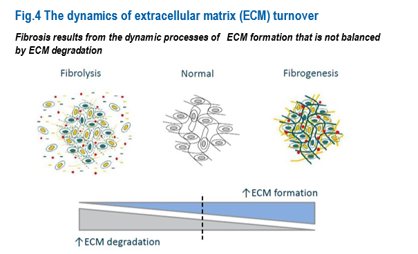
Eine Organfibrose ist das Ergebnis eines dynamischen und kontinuierlichen Prozesses der Fibrogenese (Fibroseprogression) oder Fibrolyse (Fibroseregression) (Abbildung 4).

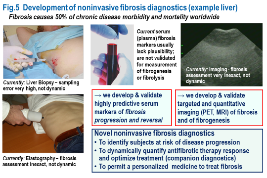
Zur Bestimmung der Schwere (des Stadiums) einer Fibrose werden mehrere nichtinvasive (nicht auf einer Gewebe-Biopsie basierende) Methoden eingesetzt. Derzeit kann jedoch keine dieser Methoden das Fibrosestadium am lebenden Patienten in vivo genau quantifizieren. Selbst die invasive Biopsie ist aufgrund ihrer geringen Größe und des damit verbundenen großen Stichprobenfehlers bei Heterogenität der Fibrose in den Geweben ungenau. Abbildung 5 zeigt die derzeitig verfügbaren in die in Entwicklung befindlichen Technologien.

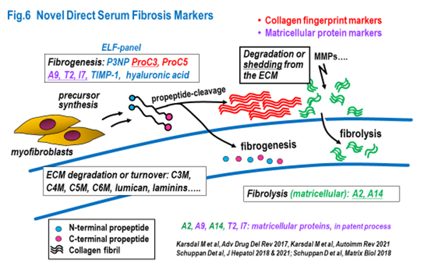
ImmuneNTech legt besonderen Wert auf die Entwicklung und Validierung serologischer Marker der Fibrogenese und Fibrolyse. Solche Marker werden als begleitende Biomarker unverzichtbar sein, um den Schweregrad der Fibrose und ein frühes Ansprechen auf eine antifibrotische Therapie vorherzusagen. Der quantitative Charakter dieser Fibrosemarker erlaubt ein Monitoring der Wirksamkeit antifibrotischer Therapien. Außerdem wird damit die Prüfung von Kombinationstherapien sowie eine personalisierte medikamentöse Behandlung ermöglicht (Abbildung 6).

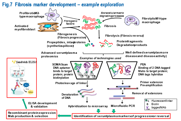
Abbildung 7 veranschaulicht die Proteom-Strategien zur Ermittlung und Validierung von Serum-(Plasma-)Biomarkern für fibrotische (Leber-)Erkrankungen, aber auch für fibrotische, entzündliche und Autoimmunerkrankungen anderer Organe, einschließlich Lunge, Nieren und Darm.

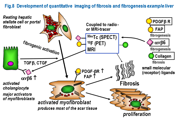
Die Wissenschaftler von ImmuneNTech entwickeln und validieren Strategien für eine gezielte quantitative Bildgebung der Fibrose und Fibrogenese (Abbildung 8).

  1. Bel Lassen P, Nori N, Bedossa P, Genser L, Aron-Wisnewsky J, Poitou C, Surabattula R, Nielsen MJ, Karsdal MA, Leeming DJ, Schuppan D*, Clément K*. Fibrogenesis marker PRO-C3 is higher in advanced liver fibrosis and improves in patients undergoing bariatric surgery. J Clin Endocrinol Metab. 2021:dgab897. doi: 10.1210/clinem/dgab897. Epub ahead of print. PMID: 34905051.
  2. Schuppan D, Myneni S, Surabattula R. Liquid biomarkers for fibrotic NASH – progress in a complex field. J Hepatol. 2021:S0168-827802173-5.
  3. Karsdal MA, Kraus VB, Shevell D, Bay-Jensen AC, Schattenberg J, Rambabu Surabattula R, Schuppan D. Profiling and targeting connective tissue remodeling in autoimmunity – A novel paradigm for diagnosing and treating chronic diseases. Autoimmun Rev. 2021;20:102706.
  4. Leeming DJ, Nielsen SH, Vongsuvanh R, Uchila P, Nielsen MJ, Reese-Petersen AL, van der Poorten D, Eslam M, Schuppan D, Karsdal MA, George J. Endotrophin, a pro-peptide of Type VI collagen, is a biomarker of survival in cirrhotic patients with hepatocellular carcinoma. Hepat Oncol. 2020;8(2):HEP32.
  5. Zimmermann A, Darstein F, Hoppe-Lotichius M, Toenges G, Lautem A, Abel F, Schad A, Mittler J, Vollmar J, Grimm D, Lang H, Galle PR, Zimmermann T, Schuppan D. Cirrhosis risk score of the donor organ predicts early fibrosis progression after liver transplantation. J Gastrointestin Liver Dis. 2019;28:53-61.
  6. Labenz C, Huber Y, Kalliga E, Nagel M, Ruckes C, Straub BK, Galle PR, Wörns MA, Anstee QM, Schuppan D, Schattenberg JM. Predictors of advanced fibrosis in non-cirrhotic non-alcoholic fatty liver disease in Germany. Aliment Pharmacol Ther. 2018;48):1109-1116.
  7. Schuppan D, Surabattula R, Wang XY. Determinants of fibrosis progression and regression in NASH. J Hepatol. 2018;68:238-250.
  8. Tanwar S, Trembling PM, Hogan BJ, Srivastava A, Parkes J, Harris S, Grant P, Nastouli E, Ocker M, Wehr K, Herold C, Neureiter D, Schuppan D, Rosenberg WM. Noninvasive markers of liver fibrosis: on-treatment changes of serum markers predict the outcome of antifibrotic therapy. Eur J Gastroenterol Hepatol. 201;29:289-296.
  9. Karsdal MA, Henriksen K, Nielsen MJ, Byrjalsen I, Leeming DJ, Gardner S, Goodman Z, Patel K, Krag A, Christiansen C, Schuppan D. Fibrogenesis assessed by serological type III collagen formation identifies patients with progressive liver fibrosis and responders to a potential antifibrotic therapy. Am J Physiol Gastrointest Liver Physiol. 2016;311:G1009-G1017.
  10. Nielsen MJ, Karsdal MA, Kazankov K, Grønbaek H, Krag A, Leeming DJ, Schuppan D, George J. Fibrosis is not just fibrosis – basement membrane modelling and collagen metabolism differs between hepatitis B- and C-induced injury. Aliment Pharmacol Ther. 2016;44:1242-1252.
  11. Tanwar S, Trembling PM, Hogan BJ, Parkes J, Harris S, Grant P, Nastouli E, Ocker M, Wehr K, Herold C, Neureiter D, Schuppan D, Rosenberg WM. Biomarkers of Hepatic Fibrosis in Chronic Hepatitis C: A Comparison of 10 Biomarkers Using 2 Different Assays for Hyaluronic Acid. J Clin Gastroenterol. 2017;51:268-277.
  12. Bernuth S, Yagmur E, Schuppan D, Sprinzl MF, Zimmermann A, Schad A, Kittner JM, Weyer V, Knapstein J, Schattenberg JM, Wörns MA, Galle PR, Zimmermann T. Early changes in dynamic biomarkers of liver fibrosis in hepatitis C virus- infected patients treated with sofosbuvir. Dig Liver Dis. 2016;48:291-7.
  13. Karsdal MA, Genovese F, Madsen EA, Manon-Jensen T, Schuppan D. Collagen and tissue turnover as a function of age: Implications for fibrosis. J Hepatol. 2016;64:103-9.
  14. Jia J, Hou J, Ding H, Chen G, Xie Q, Wang Y, Zeng M, Zhao J, Wang T, Hu X, Schuppan D. Transient elastography compared to serum markers to predict liver fibrosis in a cohort of Chinese patients with chronic hepatitis B. J Gastroenterol Hepatol. 2015;30:756-62.
  15. Nielsen MJ, Veidal SS, Karsdal MA, Ørsnes-Leeming DJ, Vainer B, Gardner SD, Hamatake R, Goodman ZD, Schuppan D, Patel K. Plasma Pro-C3 (N-terminal type III collagen propeptide) predicts fibrosis progression in patients with chronic hepatitis C. Liver Int. 2015;35:429-37.
  16. Kwong GA, von Maltzahn G, Murugappan G, Abudayyeh O, Mo S, Papayannopoulos IA, Sverdlov DY, Liu SB, Warren AD, Popov Y, Schuppan D, Bhatia SN. Mass-encoded synthetic biomarkers for multiplexed urinary monitoring of disease. Nat Biotechnol. 2013;31:63-70.

Leber-, Kolon- und Lungenkrebs

Eines der wesentlichen Ziele von ImmuneNTech ist die Entwicklung und klinische Translation von effizienten Therapien für diese Krebstypen.

Kontakt

Haben wir Ihr Interesse geweckt? Kontaktieren Sie uns gerne telefonisch oder über das Kontaktformular.

Zurück nach oben